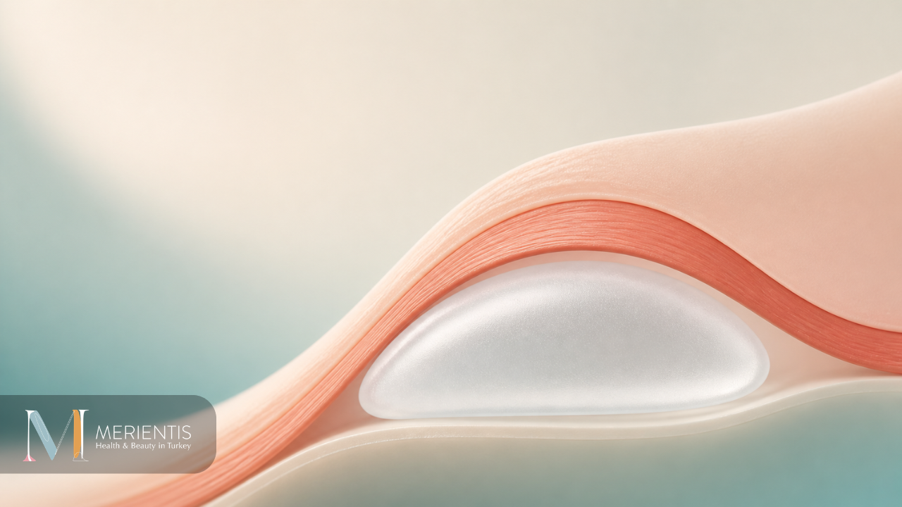
Most breast implants today are placed partially under the pectoral muscle using the dual plane technique. The muscle covers the upper part of the implant, softening the transition that creates a round, obvious look in thin patients, while the lower part sits under breast tissue to allow natural shape and movement.
“Where exactly is the implant hidden to make the result look natural?”
This question comes up in almost every consultation, and it is a better question than most patients realise. The implant itself matters less than what covers it. Two women can receive the same implant and end up with completely different results — because the pocket the implant sits in changes how much of its edge the eye can see.
This article explains the three placement planes, why the dual plane became the default choice for most surgeons, what the published evidence actually supports, and what the decision means for recovery — particularly if you are travelling for surgery.
The three placement planes
An implant can be positioned in one of three main layers of the chest wall. Each changes how much tissue lies between the implant and the skin.
| Plane | Where the implant sits | Main characteristic |
|---|---|---|
| Subglandular (over the muscle) | Directly beneath breast tissue, on top of the pectoral muscle | Simplest dissection, least postoperative discomfort, least coverage |
| Subfascial | Beneath the thin fascia covering the muscle, still above the muscle itself | A middle option; the fascia adds a small amount of camouflage |
| Submuscular / dual plane (under the muscle) | Upper portion under the pectoral muscle, lower portion under breast tissue | Most upper pole coverage; the current default in most practices |
These are not equally common. In a survey of 1,067 plastic surgeons in the United States, partial submuscular or dual plane placement accounted for 79.5% of cases, complete submuscular for 12.7%, subglandular for 5.4%, and subfascial for 2.4%. Subfascial placement is more frequent in Europe and South America than in the United States.
Why thin patients get the “ball on the chest” look
This is the part that explains the whole decision, and it has a measurable clinical basis rather than an aesthetic opinion.
Surgeons assess soft tissue thickness at the upper pole of the breast using a pinch test. Skin and tissue are gently pinched at the top of the breast and the thickness measured. When the upper pole pinch measures less than roughly 2 cm, there is not enough natural tissue to disguise the top edge of an implant.
Place an implant directly under thin breast tissue and the eye sees exactly where the device begins. The result is a visible step-off at the top of the breast — an abrupt convex curve rather than a gentle slope. That is the “round”, “stuck-on” or “ball-like” appearance patients describe, and it is not caused by the implant being too large. It is caused by insufficient coverage.
The pectoral muscle solves this specific problem. When the upper portion of the implant sits beneath the muscle, the muscle compresses and blurs the upper edge of the device, so the transition from chest to breast becomes gradual rather than sudden.
This is why the plane decision matters most in slim patients with little natural breast tissue — precisely the group most likely to want augmentation and most at risk of an obviously operated result.
What “dual plane” actually means
Complete submuscular placement — the entire implant beneath the muscle — has a drawback. The muscle holds the implant high and restricts the lower pole, so the breast can look flat at the bottom and the implant may sit above the natural breast fold.
The dual plane approach was developed to solve this. The pectoral muscle covers the upper portion of the implant, while the lower portion sits directly beneath breast tissue. The upper pole gets the coverage it needs; the lower pole gets the freedom to fill out and drape naturally.
The extent to which the muscle is released varies according to breast anatomy — how much tissue is present, how much the breast has descended, and where the natural fold sits. This adjustment is what makes the technique adaptable rather than a single fixed manoeuvre, and it is decided during the operation rather than in advance.
What the evidence actually supports
The comparison between planes is often presented as though one is simply better. The published data supports a more specific conclusion.
Capsular contracture
This is the outcome with the clearest and most consistent evidence. A systematic review and meta-analysis of 24 studies found that subpectoral placement was associated with significantly lower capsular contracture rates than prepectoral placement (OR 0.35; 95% CI 0.25-0.50; P < .00001). Other pairwise analyses have reported roughly a three-fold higher risk with subglandular compared with submuscular placement.
A 2024 systematic review comparing subfascial with subglandular placement found the subfascial plane associated with lower rates of capsular contracture, rippling and haematoma — though the authors noted all included studies carried a high risk of bias.
The pattern across all of this is consistent: planes with more tissue coverage show lower capsular contracture risk.
Rippling and visible implant edges
Rippling — visible surface irregularity where the implant edge shows through the skin — occurs more often with less coverage. Here, though, an honest caveat is required.
Much of the comparative data on rippling and animation deformity comes from breast reconstruction studies rather than cosmetic augmentation. In reconstruction, prepectoral placement showed higher rippling rates (OR 2.21; 95% CI 1.52-3.21). Reconstruction patients have thinner tissue coverage than augmentation patients by definition, so these figures cannot be transferred directly.
Capsular contracture remains the only outcome for which augmentation-specific meta-analytic evidence is available. Anyone quoting precise rippling percentages for cosmetic augmentation is going beyond what the literature currently supports.
The trade-off: animation deformity
Placing an implant under the muscle introduces a specific issue that does not occur above it.
When the pectoral muscle contracts — pushing, lifting, certain gym movements — it can move the implant beneath it, causing temporary distortion or flattening of the breast. This is called animation deformity. In reconstruction data, submuscular placement showed significantly higher rates than prepectoral placement (OR 0.09; 95% CI 0.03-0.25 favouring prepectoral).
For most patients the effect is mild and only visible during deliberate muscle contraction. For competitive athletes, bodybuilders and those whose work involves heavy repetitive upper body effort, it can matter more, and the plane decision may reasonably shift.
Submuscular placement also involves more early discomfort. Muscle has been lifted and partly released, and it responds accordingly — with spasm and soreness during the first days.
What the decision means for recovery and travel
If you are travelling for surgery, the plane affects your first week more than most consultations mention.
Submuscular and dual plane placement typically involve more early discomfort than subglandular placement, particularly with movements that engage the chest — reaching up, pushing a door, lifting hand luggage into an overhead locker. Muscle spasm in the first days is common and settles as the muscle adapts.
Two practical implications follow. First, arrangements involving heavy lifting during the return journey should be planned around this; assistance with luggage is worth organising in advance. Second, the timing of the return flight is determined by your recovery, not by the plane alone, and should be confirmed by the treating surgeon rather than assumed at booking.
None of this argues against submuscular placement. The early discomfort is temporary; the coverage is permanent. But knowing what the first week involves makes it easier to plan realistically.
Which plane suits whom?
| Situation | Consideration |
|---|---|
| Thin patient, upper pole pinch under ~2 cm | Coverage is the priority; submuscular or dual plane usually favoured |
| Good existing breast tissue | More options open; natural tissue already provides camouflage |
| Competitive athlete or heavy upper body work | Animation deformity weighs more heavily in the decision |
| Some degree of sagging present | The fold position and tissue distribution influence pocket choice; a lift may also be discussed |
| Priority on lowest capsular contracture risk | Evidence favours planes with greater coverage |
No single plane is correct for everyone, and this is genuinely an anatomical decision rather than a preference. Tissue thickness, breast shape, fold position, implant choice and lifestyle all feed into it.
Questions worth asking before surgery
- Which plane is being recommended for me, and what specifically about my anatomy leads to that choice?
- What was my upper pole pinch measurement?
- Given my tissue thickness, how visible is the implant edge likely to be?
- How much animation is realistic for me with this plane?
- How will the first week feel, and when can I lift and reach normally?
- If I am unhappy with the result later, what would revision involve?
A clear, anatomy-specific answer to the first two questions is a good sign. A generic answer is worth probing further.
In summary
The plane decision is about coverage, and coverage is what separates a natural result from an obvious one — especially in slim patients where thin upper pole tissue cannot disguise an implant edge on its own. The dual plane technique became the most widely used approach because it addresses that problem without holding the lower breast flat.
The evidence most firmly supports lower capsular contracture risk with greater tissue coverage. The main trade-off is animation deformity and more early discomfort. Which balance suits you depends on measurements taken during examination, not on what worked for someone else.
If you would like your own anatomy assessed and the reasoning behind a plane recommendation explained, you can arrange a consultation to discuss what is realistic in your case. You may also find whether breast implants feel natural a useful companion to this article, and how breast lift scars fade relevant if a lift has also been mentioned to you.
Frequently Asked Questions
Are breast implants better under or over the muscle?
Neither is universally better. Placement under the muscle provides more upper pole coverage and is associated with lower capsular contracture rates, while placement over the muscle avoids animation deformity and involves less early discomfort. The right choice depends on tissue thickness, breast shape and lifestyle.
What is dual plane breast augmentation?
Dual plane placement positions the upper portion of the implant beneath the pectoral muscle and the lower portion beneath breast tissue. This combines upper pole coverage with natural lower pole shape. It is the most commonly used approach, accounting for around 79.5% of cases in a survey of 1,067 US plastic surgeons.
Why do some implants look round and obvious?
Usually because there is not enough tissue covering the upper edge of the implant, creating a visible step-off rather than a gradual slope. When the upper pole pinch test measures under roughly 2 cm, muscle coverage is generally needed to soften that transition.
Does under the muscle placement hurt more?
The early recovery typically involves more discomfort, because the muscle has been lifted and partially released. Muscle spasm during the first days is common and settles as the muscle adapts. The difference is temporary.
What is animation deformity?
It is temporary distortion of the breast when the pectoral muscle contracts, which can occur when an implant sits beneath the muscle. For most patients it is mild and only visible during deliberate contraction, but it can matter more for athletes and those doing heavy upper body work.
Which plane has the lowest risk of capsular contracture?
Evidence favours planes with greater tissue coverage. A meta-analysis of 24 studies found subpectoral placement associated with significantly lower capsular contracture rates than prepectoral placement (OR 0.35; 95% CI 0.25-0.50).
Can the plane be changed later?
Changing the pocket is possible but constitutes revision surgery with its own considerations and recovery. This is one reason the initial decision is worth understanding rather than accepting without discussion.
Does the plane affect when I can fly home?
Return travel timing is determined by your overall recovery and confirmed by the treating surgeon. Submuscular placement involves more early discomfort with reaching and lifting, so arranging assistance with luggage is worth planning in advance.
Medically reviewed by Dr. Ahmet Kaplan, Specialist in Plastic, Reconstructive and Aesthetic Surgery (EBOPRAS, 2022), Istanbul.
Merientis Health is a health tourism facilitator and is not a healthcare facility. All medical procedures are performed by the treating physician at the relevant licensed healthcare facility. This content is for informational purposes only and does not replace medical examination, diagnosis or treatment. Individual results vary. Please consult a qualified physician regarding your own situation.
References
- Capsular Contracture After Breast Augmentation: A Systematic Review and Meta-Analysis. Aesthetic Surgery Journal Open Forum, 2025.
- Outcomes in Subfascial Versus Subglandular Planes in Breast Augmentation: A Systematic Review and Meta-analysis. Aesthetic Surgery Journal 2024;44(9):NP639.
- Implant-based breast reconstruction — a systematic review and meta-analysis of prepectoral versus submuscular implant placement. Annals of Breast Surgery.
- Hidalgo DA. Survey of implant pocket selection among 1,067 plastic surgeons.